At Dakota Eye Care Associates our motto is “Leaders in eye care today and tomorrow”. We stay true to this by continually updating our technology with new instrumentation and new products.
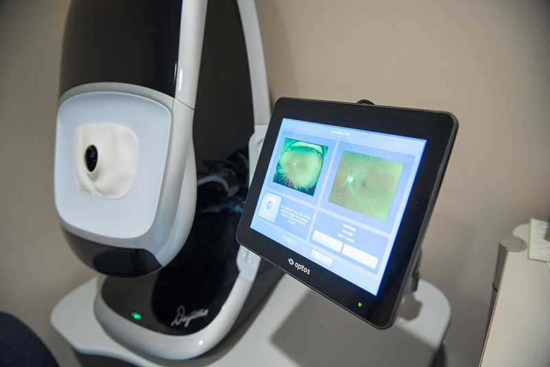
Optomap Retinal Photography:
This provides a 200-degree panoramic, digital image of the retina to aid in the diagnosis of retina disorders such as macular degeneration, diabetic retinopathy and hypertensive retinopathy. Learn more at optos.com

Optomap Retinal Photography:
This provides a 200-degree panoramic, digital image of the retina to aid in the diagnosis of retina disorders such as macular degeneration, diabetic retinopathy and hypertensive retinopathy. Learn more at optos.com
FDT (Frequency Doubling Technology)
A test used to screen for visual field loss. Highly specific in detecting changes from glaucoma, macular disorders and neurological changes.
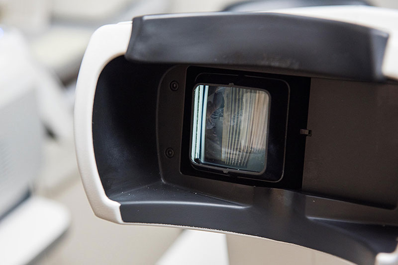

FDT (Frequency Doubling Technology)
A test used to screen for visual field loss. Highly specific in detecting changes from glaucoma, macular disorders and neurological changes.
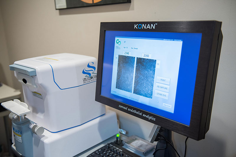
Specular Microscope:
A photographic technique used to detect changes within the corneal endothelium. This is used to detect many corneal diseases and impacts of contact lens wear.

Specular Microscope:
A photographic technique used to detect changes within the corneal endothelium. This is used to detect many corneal diseases and impacts of contact lens wear.
OCT:
A noninvasive technique used to monitor the health of the 10 different retinal layers and optic nerve head. This tool is useful in the diagnosis and management of macular degeneration and glaucoma.
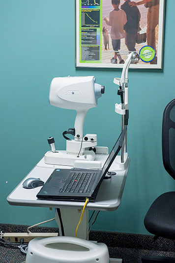

OCT:
A noninvasive technique used to monitor the health of the 10 different retinal layers and optic nerve head. This tool is useful in the diagnosis and management of macular degeneration and glaucoma.
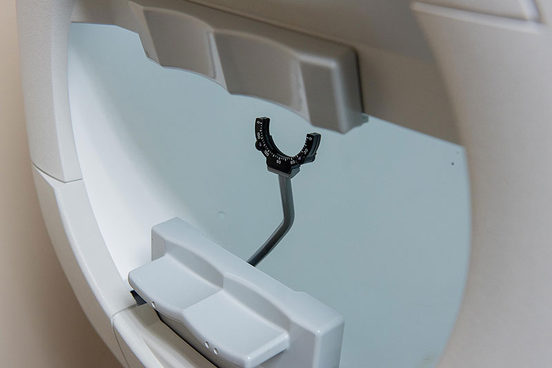
Humphrey Visual Field:
An instrument used to map out a patient’s visual field. This is a more in-depth test to for detection and management of glaucoma, macular and neurological changes.

Humphrey Visual Field:
An instrument used to map out a patient’s visual field. This is a more in-depth test to for detection and management of glaucoma, macular and neurological changes.
VEP/ERG:
Detects the rate and change at which information travels from the eye to the brain. This is highly specific for diseases that damage the optic nerve (Multiple sclerosis, glaucoma) and the retina.
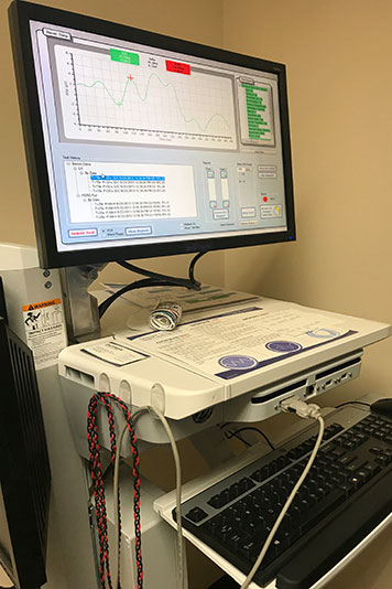

VEP/ERG:
Detects the rate and change at which information travels from the eye to the brain. This is highly specific for diseases that damage the optic nerve (Multiple sclerosis, glaucoma) and the retina.
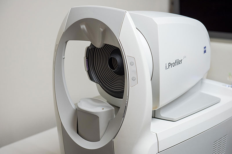
Corneal Topography:
Maps the contours of the cornea and detects irregularities from contact lens wear and corneal diseases such as keratoconus.

Corneal Topography:
Maps the contours of the cornea and detects irregularities from contact lens wear and corneal diseases such as keratoconus.
